The double helical structure of DNA was proposed by James Watson and Francis Crick in 1953 (Noble Prize, 1962). The structure of DNA double helix is comparable to a twisted ladder as shown in Figure 5.3.
The salient features of Watson and Crick model of DNA are given below.
DNA molecule consists of two helical polynucleotide chains, which are coiled around a common axis in the form of a right-handed double helix. Such double helices cannot be pulled apart and can be separated only by an unwinding process.
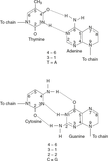
Figure 5.3 Complementary Base Pairing in DNA
They are called plectonemic coils, that is, coils that are interlocked about the same axis.
The two strands are antiparallel, that is, one strand runs from 5′ to 3′ direction, while the other from 3′ to 5′ direction. This is comparable to two parallel adjacent roads carrying traffic in opposite direction.
The width (or diametre) of a double helix is 20 Å(2 nm).
Each turn of the helix is 34 Å (3.4 nm) with 10 pairs of nucleotides, with each pair at a distance of about 3.4 Å.
Each DNA strand has a hydrophilic deoxyribose phosphate backbone (3′-5′−phosphodiester bond) facing outwards, while the hydrophobic bases are stacked inside.
The two nucleotides are complementary to each other due to the base pairing.
The two strands are held together by hydrogen bonds formed by complementary base pairs. The A—T pair has two hydrogen bonds while G—C pair has three hydrogen bonds. The G≡C is stronger by about 50% than A=T.
The hydrogen bonds are formed between purines and pyrimidines only. If two purines face each other, they would not fit into the allowable space. And two pyrimidines would be too far to form hydrogen bonds. The only base arrangement possible in DNA structure, from special considerations, is A—T, T—A, G—C, and C—G.
Chargaff’s rule explains that the DNA base pairs are complementary to each other. So, the number of adenine equals to that of thymine (A = T) and the number of guanine is equal to that of cytosine (G = C).
The genetic information resides on one of the two strands known as template strand or sense strand. The opposite strand is antisense strand. The double helix has major grooves and minor grooves along the phosphodiester backbone. Proteins interact with DNA at these grooves, without disrupting the base pairs and double helix. The diagrammatic representation of Watson–Crick model of double helical structure of DNA is given in Figure 5.4.
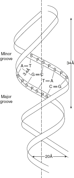
Figure 5.4 Watson–Crick Model of Double Helical Structure of DNA
Leave a Reply